Correlati neuropsicologici e di neuroimaging del maltrattamento e dell’abuso infantili

Titolo Rivista MALTRATTAMENTO E ABUSO ALL’INFANZIA
Autori/Curatori Marco Cavallo, Arianna Signorino
Anno di pubblicazione 2015 Fascicolo 2015/3 Lingua Italiano
Numero pagine 23 P. 11-33 Dimensione file 130 KB
DOI 10.3280/MAL2015-003002
Il DOI è il codice a barre della proprietà intellettuale: per saperne di più clicca qui

Qui sotto puoi vedere in anteprima la prima pagina di questo articolo.

Se questo articolo ti interessa, lo puoi acquistare (e scaricare in formato pdf) seguendo le facili indicazioni per acquistare il download credit. Acquista Download Credits per scaricare questo Articolo in formato PDF
Anteprima articolo

FrancoAngeli è membro della Publishers International Linking Association, Inc (PILA)associazione indipendente e non profit per facilitare (attraverso i servizi tecnologici implementati da CrossRef.org) l’accesso degli studiosi ai contenuti digitali nelle pubblicazioni professionali e scientifiche

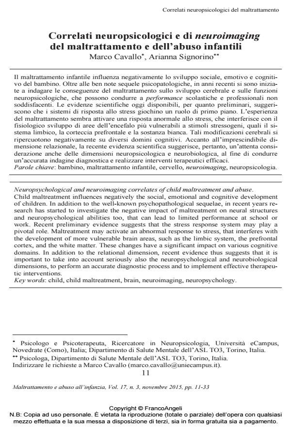
Il maltrattamento infantile influenza negativamente lo sviluppo sociale, emotivo e cognitivo del bambino. Oltre alle ben note sequele psicopatologiche, in anni recenti si sono iniziate a indagare le conseguenze del maltrattamento sullo sviluppo cerebrale e sulle funzioni neuropsicologiche, che possono condurre a performance scolastiche e professionali non soddisfacenti. Le evidenze scientifiche oggi disponibili, per quanto preliminari, suggeriscono che i sistemi di risposta allo stress giochino un ruolo di primo piano. L’esperienza del maltrattamento sembra attivare una risposta anormale allo stress, che interferisce con il fisiologico sviluppo di aree dell’encefalo più vulnerabili a stimoli stressogeni, quali il sistema limbico, la corteccia prefrontale e la sostanza bianca. Tali modificazioni cerebrali si ripercuotono negativamente su diversi domini cognitivi. Accanto all’imprescindibile dimensione relazionale, la recente evidenza scientifica suggerisce, pertanto, un’attenta considerazione anche delle dimensioni neuropsicologica e neurobiologica, al fine di condurre un’accurata indagine diagnostica e realizzare interventi terapeutici efficaci.;

Keywords:Bambino, maltrattamento infantile, cervello, neuroimaging, neuropsicologia

  1. Alink, L. R., Cicchetti, D., Kim, J., & Rogosch, F. A. (2012). Longitudinal associations among child maltreatment, social functioning, and cortisol regulation. Developmental Psychology, 48, 1, 224-236, DOI: 10.1037/a0024892
  2. Anderson, P. (2002). Assessment and development of executive function (EF) during childhood. Child Neuropsychology, 8, 2, 71-82, DOI: 10.1076/chin.8.2.71.8724
  3. Arnsten, A. F. T. (2009). Stress signalling pathways that impair prefrontal cortex structure and function. Nature Reviews Neuroscience, 10, 6, 410-422, DOI: 10.1038/nrn2648
  4. Attili, G., Di Pentima, L., & Toni, A. (2013). Abuso, trascuratezza e il controllo degli eventi: il paradosso dell’attaccamento. Maltrattamento e abuso all’infanzia, 15, 3, 89-108, DOI: 10.3280/MAL2013-003005
  5. Bauer, P. M., Hanson, J. L., Pierson, R. K., Davidson, R. J., & Pollak, S. D. (2009). Cerebellar volume and cognitive functioning in children who experienced early deprivation. Biological Psychiatry, 66, 12, 1100-1106, DOI: 10.1016/j.biopsych.2009.06.014
  6. Beers, S. R., & De Bellis, M. D. (2002). Neuropsychological function in children with maltreatment-related posttraumatic stress disorder. American Journal of Psychiatry, 159, 3, 483-486, DOI: 10.1176/appi.ajp.159.3.483
  7. Bick, J., Zhu, T., Stamoulis, C., Fox, N. A., Zeanah, C., & Nelson, C. A. (2015). A randomized clinical trial of foster care as an intervention for early istituzionalization: long term improvements in white matter microstructure. JAMA Pediatrics, 169, 3, 211-219, DOI: 10.1001/jamapediatrics.2014.3212
  8. Bremner, J. D., Randall, P., Vermetten, E., Staib, L., Bronen, R. A., Mazuro, C., Mazure, C., Capelli, S., McCarthy, G., Innis, R. B., & Charney, D. S. (1997).
  9. Magnetic resonance imaging-based measurement of hippocampal volume in posttraumatic stress disorder related to childhood physical and sexual abuse. A preliminary report. Biological Psychiatry, 41, 1, 23-32, DOI: 10.1016/s0006-3223(96)00162-x
  10. Bruce, J., Fisher, P. A., Pears, K. C., & Levine, S. (2009). Morning cortisol levels in preschool-aged foster children: Differential effects of maltreatment type. Developmental Psychobiology, 51, 1, 14-23, DOI: 10.1002/dev.20333
  11. Camisasca, E. (2009). Traiettorie di internalizzazione ed esternalizzazione in bambini maltrattati: il ruolo dell’attaccamento. Maltrattamento e abuso all’infanzia, 3, 65-82. DOI: 10.3280/MAL2009-003007
  12. Camisasca, E., Miragoli, S., & Di Blasio, P. (2014). La disorganizzazione dell’attaccamento spiega i sintomi post-traumatici nei bambini vittime di violenza? Maltrattamento e abuso all’infanzia, 16, 2, 35-55. DOI: 10.3280/MAL2014-002003
  13. Carrion, V. G., Weems, C. F., Eliez, S., Patwardhan, A., Brown, W., Ray, R. D., & Reiss, A. L. (2001). Attenuation of frontal asymmetry in pediatric posttraumatic stress disorder. Biological Psychiatry, 50, 12, 943-951, DOI: 10.1016/s0006-3223(01)01218-5
  14. Carrion, V. G., Weems, C. F., Ray, R. D., Glaser, B., Hessl, D., & Reiss. A. L. (2002). Diurnal salivary cortisol in pediatric posttraumatic stress disorder. Biological Psychiatry, 51, 7, 575-582, DOI: 10.1016/s0006-3223(01)01310-5
  15. Carrion, V. G., Weems, C. F., Watson, C., Eliez, S., Menon, V., & Reiss, A. L. (2009). Converging evidence for abnormalities of the prefrontal cortex and evaluation of midsagittal structures in pediatric posttraumatic stress disorder: An MRI study. Psychiatry Research Neuroimaging, 172, 3, 226-234, DOI: 10.1016/j.pscychresns.2008.07.008
  16. Cigala, A., & Mori, A. (2012). Le competenze emotive in bambini con storia di maltrattamento: cosa ci dice la ricerca? Maltrattamento e abuso all’infanzia, 14, 1, 11-24, DOI: 10.3280/MAL2012-001002
  17. Dannlowski, U., Stuhrmann, A., Beutelmann, V., Zwanzger, P., Lenzen, T., Grotegerd, D., Domschke, K., Hohoff, C., Ohrmann, P., Bauer, J., Lindner, C., Postert, C., Konrad, C., Arolt, V., Heindel, W., Suslow, T., & Harald, K. (2012a). Limbic scars: Long-term consequences of childhood maltreatment revealed by functional and structural magnetic resonance imaging. Biological Psychiatry, 71, 4, 286-293, DOI: 10.1016/j.biopsych.2011.10.021
  18. Dannlowski, U., Kugel, H., Huber, F., Stuhrmann, A., Redlich, R., Grotegerd, D., Dohm, K., Sehlmeyer, C., Konrad, C., Baune, B. T., Arolt, V., Heindel, W., Zwitserlood, P., & Suslow, T. (2012b). Childhood maltreatment is associated with an automatic negative emotion processing bias in the amygdala. Human Brain Mapping, 34, 11, 2899-2909 DOI: 10.1002/hbm.22112
  19. De Bellis, M. D. (2001). Developmental traumatology: The psychobiological development of maltreated children and its implications for research, treatment and policy. Developmental Psychopathology, 13, 3, 539-564, DOI: 10.1017/s0954579401003078
  20. De Bellis, M. D. (2003). The neurobiology of post-traumatic-stress disorder across the life cycle. In J. C. Soares & S. Gershon (Eds.) The handbook of medical psychiatry (pp. 449-466). New York: Marcel Dekker, DOI: 10.3109/9780203912485-32
  21. De Bellis, M. D., Baum, A., Birmaher, B., Keshavan, M., Eccard, C. H., Boring, A. M., Jenkins, F. J., & Ryan, N. D. (1999). Developmental traumatology part I: Biological stress systems. Biological Psychiatry, 45, 10, 1259-1270, DOI: 10.1016/s0006-3223(99)00044-x
  22. De Bellis, M. D., Keshavan, M. S., Shifflett, H., Iyengar, S., Beers, S. R., Hall, J., & Moritz, G. (2002). Brain structure in pediatric maltreatment-related posttraumatic stress disorder: A sociodemographically matched study. Biological Psychiatry, 52, 11, 1066-1078, DOI: 10.1016/s0006-3223(02)01459-2
  23. De Bellis, M. D., & Kuchibhatla, M. (2006). Cerebellar volumes in pediatric maltreatment-related posttraumatic stress disorder. Biological Psychiatry, 60, 7, 697-703, DOI: 10.1016/j.biopsych.2006.04.035
  24. De Bellis, M. D., Hooper, S. R., Spratt, E. G., & Woolley, D. P. (2009). Neuropsychological findings in childhood neglect and their relationships to pediatric PTSD. Journal of International Neuropsychological Society, 15, 6, 868-878, DOI: 10.1017/s1355617709990464
  25. De Bellis, M. D., & Hooper, S. R. (2012). Neural substrates for processing taskirrelevant emotional distracters in maltreated adolescents with depressive disorders: A pilot study. Journal of the International Traumatic Stress Society, 25, 2, 198-202, DOI: 10.1002/jts.21682
  26. De Bellis, M. D., Woolley, D. P., & Hooper, S. R. (2013). Neuropsychological findings in pediatric maltreatment: Relationship of PTSD, dissociative symptoms, and abuse/neglect indices to neurocognitive outcomes. Child Maltreatment, 18, 3, 171-183, DOI: 10.1177/1077559513497420
  27. De Brito, S., Viding, E., Sebastian, C. L., Kelly, P. A., Mechelli, A., Maris, H., & McCrory, E. J. (2012). Reduced orbitofrontal and temporal grey matter in a community sample of maltreated children. Journal of Child Psychology and Psychiatry, 54, 1, 105-112, DOI: 10.1111/j.1469-7610.2012.02597.x.
  28. Di Blasio, P., Camisasca, E., & Procaccia, R. (2007). Fattori di mediazione dell’esperienza traumatica nei bambini maltrattati. Maltrattamento e abuso all’infanzia, 2, 33-59.
  29. Eluvathingal, T. J., Chugani, H. T., Behen, M. E., Juhàsz, C., Muzik, O., Maqbool, M., Chugani, D. C., & Makki, M. (2006). Abnormal brain connectivity in children after early severe socioemotional deprivation: A diffusion tensor imaging study. Pediatrics, 117, 6, 2093-2100, DOI: 10.1542/peds.2005-1727
  30. Gazzaniga, M. S., Ivry, R. B., & Mangun, G. R. (2002). Sviluppo e plasticità. In M. S. Gazzaniga, R. B. Ivry, & G. R. Mangun (Eds.), Neuroscienze cognitive. Bologna: Zanichelli.
  31. Gordon, J. A., & Hen, R. (2004). The serotonergic system and anxiety. Neuromolecular Medicine, 5, 1, 27-40, DOI: 10.1385/nmm:5:1:027
  32. Govindan, R. M., Behen, M. E., Helder, E., Makki, M. I., & Chugani, H. T. (2009). Altered water diffusivity in cortical association tracts in children with early deprivation identified with Tract-Based Spatial Statistics (TBSS). Cerebral Cortex, 20, 3, 561-569, DOI: 10.1093/cercor/bhp122
  33. Gunnar, M. R., Brodersen, L., Krueger, K., & Rigatuso, J. (1996). Dampening of adrenocortical responses during infancy: Normative changes and individual differences. Child Development, 67, 3, 877-889, DOI: 10.2307/1131867
  34. Gunnar, M. R., Morison, S. J., Chisholm, K., & Schuder, M. (2001). Salivary cortisol levels in children adopted from Romanian orphanages. Development & Psychopathology, 13, 3, 611-628, DOI: 10.1017/s095457940100311x
  35. Gunnar, M. R., & Quevedo, K. M. (2007). Early care experiences and HPA axis regulation in children: A mechanism for later trauma vulnerability. Progress in Brain Research, 167, 137-149, DOI: 10.1016/s0079-6123(07)67010-1
  36. Hanson, J. L, Adluru, N., Chung, M. K., Alexander, A. L., Davidson, R. J., & Pollak, S. D. (2013). Early neglect is associated with alterations in white matter integrity and cognitive functioning. Child Development, 84, 5, 1566-1578, DOI: 10.1111/cdev.12069
  37. Hart, J., Gunnar, M., & Cicchetti, D. (1996) Altered neuroendocrine activity in maltreated children related to symptoms of depression. Development and Psychopathology, 8, 1, 201-214, DOI: 10.1017/s0954579400007045
  38. Hill, M. N., Hillard, C. J., & McEwen, B. S. (2011). Alterations in corticolimbic dendritic morphology and emotional behaviorin cannabinoid CB1 receptordeficient mice parallel the effects of chronic stress. Cerebral Cortex, 21, 9, 2056-2064, DOI: 10.1093/cercor/bhq280
  39. Jackowski, A. P., Douglas-Palumberi, H., Jackowski, M., Win, L., Schultz, R. T., Staib, L.W., Krystal, J. H., & Kaufman, J. (2008). Corpus callosum in maltreated children with posttraumatic stress disorder: A diffusion tensor imaging study. Psychiatry Research Neuroimaging, 162, 3, 256-261, DOI: 10.1016/j.pscychresns.2007.08.006.
  40. J., & Kaufman, J. (2011). Early-life stress, corpus callosum development, hippocampal volumetrics, and anxious behavior in male nonhuman primates. Psychiatry Research Neuroimaging, 192, 1, 37-44, DOI: 10.1016/j.pscychresns.2010.11.006
  41. Kaufman, J., Birmaher, B., Perel, J., Dahl, R. E., Moreci, P., Nelson, B., & Ryan, N. (1997). The corticotropin-releasing hormone challenge in depressed abused, depressed nonabused, and normal control children. Biological Psychiatry, 42, 8, 669-679, DOI: 10.1016/s0006-3223(96)00470-2
  42. Lebel, C., Gee, M., Camicioli, R., Wieler, M., Martin, W., & Beaulieu, C. (2012).
  43. Diffusion tensor imaging of white matter tract evolution over the lifespan. NeuroImage, 60, 340-352, DOI: 10.1016/j.neuroimage.2011.11.094
  44. LeDoux, J. E. (2000). Emotion circuits in the brain. Annual Review Neuroscience, 23, 1, 155-184, DOI: 10.1146/annurev.neuro.23.1.155
  45. Lupien, S. J., McEwan, B. S., Gunnar, M. R., & Heim, C. (2009). Effects of stress throughout the lifespan on the brain, behaviour and cognition. Nature Reviews Neuroscience, 10, 6, 434-445, DOI: 10.1038/nrn2639
  46. Lyoo, I. K., Noam, G. G, Lee, C. K., Lee, H. K, Kennedy, B. P., & Renshaw, P. F. (1996). The corpus callosum and lateral ventricles in children with attentiondeficit hyperactivity disorder: A brain magnetic resonance imaging study. Biological Psychiatry, 40, 10, 1060-1063, DOI: 10.1016/s0006-3223(96)00349-6
  47. Maheu, F. S., Dozier, M., Guyer, A. E., Mandell, D., Peloso, E., Poeth, K., & Ernst, M. A. (2010). Preliminary study of medial temporal lobe function in youths with a history of caregiver deprivation and emotional neglect. Cognitive, Affective & Behavioural Neuroscience, 10, 1, 34-49, DOI: 10.3758/cabn.10.1.34
  48. McCrory, E., De Brito, S. A., & Viding, E. (2011). The impact of childhood maltreatment: A review of neurobiological and genetic factors. Frontiers in Psychiatry, 2, 1-14, DOI: 10.3389/fpsyt.2011.00048
  49. McCrory, E. J., De Brito, S. A., Kelly, A. P., Bird, G., Sebastian, C. L., Mechelli, A., Samuel, S., & Viding, E. (2013). Amygdala activation in maltreated children during pre-attentive emotional processing. The British Journal of Psychiatry, 202, 4, 269-276, DOI: 10.1192/bjp.bp.112.116624
  50. McQueen, G. M., & Frodl, T. (2010). The hippocampus in major depression: Evidence for the convergence of the bench and bedside in psychiatric research? Molecular Psychiatry, 16, 3, 252-264, DOI: 10.1038/mp.2010.80
  51. Mehta, M. A., Golembo, N. I., Nosarti, C., Colvert, M., Mota, A., Williams, S. C. K., Rutter, M., & Sonuga-Barke, E. J. S. (2009). Amygdala, hippocampal and corpus callosum size following severe early institutional deprivation: The English and Romanian Adoptees study pilot. Journal of Child Psychology and Psychiatry, 50, 8, 943-951, DOI: 10.1111/j.1469-7610.2009.02084.x
  52. Miller, D. J., Duka, T., Stimpson, C. D., Schapiro, S. J., Baze, W. B., McArthur, M. J., Fobbs, A. J., Sousa, A. M. M., Sestan, N., Wildman, D. E., Lipovich, L., Kuzawa, C. W., Hof, P. R., & Sherwood, C. C. (2012). Prolonged myelination in human neocortical evolution. Proceedings of the National Academy of Sciences of the United States of America, 109, 41, 16480-16485, DOI: 10.1073/pnas.1117943109
  53. Nikulina, V., & Widom, C. S. (2013). Child maltreatment and executive functioning in middle adulthood: A prospective examination. Neuropsychology, 27, 4, 417-427, DOI: 10.1037/a0032811
  54. OMS (1999). Report on the Consultation on Child Abuse Prevention. Geneva, 29-31 March 1999. World Health Organization, Social Change and Mental Health, Violence and Injury Prevention.
  55. OMS (2014). Global status report on violence prevention. World Health Organization.
  56. Pagani, M., & Cavallo, M. (2014). Neuroimaging in PTSD-related psychotherapies. In R. Dierckx, A. Otte, E. de Vries, & A. van Waarde (Eds.), PET and SPECT in Psychiatry (pp. 397-410). Berlin: Heidelberg Springer Verlag.
  57. Pajevic, S., & Pierpaoli, C. (1999). Color schemes to represent the orientation of anisotropic tissues from diffusion tensor data: Application to white matter fiber tract mapping in the human brain. Magnetic Resonance in Medicine, 42, 3, 526-540, DOI: 10.1002/(sici)1522-2594(199909)42:3<526::aid-mrm15>3.0.co;2-j
  58. Preis, S., Steinmetz, H., Knorr, U., & Jancke, L. (2000). Corpus callosum size in children with developmental language disorder. Cognitive Brain Research, 10, (1-2), 37-44, DOI: 10.1016/s0926-6410(00)00020-3
  59. Procaccia, R. (2011). Rischio e resilienza nei bambini maltrattati. Maltrattamento e abuso all’infanzia, 13, 3, 57-76, DOI: 10.3280/MAL2011-003004
  60. Purves, D., Augustine, G. J., Fitzpatrick, D., Katz, L. C., LaMantia, A. S., McNamara, J. O., & Williams, S. M. (2001). Le modificazioni dei circuiti nervosi in funzione dell’esperienza. In M. S. Gazzaniga, R. B. Ivry, & G. R. Mangun (Eds.), Neuroscienze cognitive. Bologna: Zanichelli.
  61. Rao, U., Chen, L. A., Bidesi, A. S., Shad, M. U., Thomas, M. A., & Hammen, C. L. (2010). Hippocampal changes associated with early-life adversity and vulnerability to depression. Biological Psychiatry, 67, 4, 357-364, DOI: 10.1016/j.biopsych.2009.10.017
  62. Rollins, N. K. (2007). Clinical applications of diffusion tensor imaging and tractography in children. Pediatric Radiology, 37, 8, 769-780, DOI: 10.1007/s00247-007-0524-z
  63. Samuelson, K. W., Krueger, C. E., Burnett, C., & Wilson, C. K. (2010). Neuropsychological functioning in children with posttraumatic stress disorder. Journal of Child Neuropsychology, 16, 2, 119-133, DOI: 10.1080/09297040903190782
  64. Schmahmann, J. D. (1991). An emerging concept. The cerebellar contribution to higher function. Archives of Neurology, 48, 11, 1178-1187, DOI: 10.1001/archneur.1991.00530230086029
  65. Schmahmann, J. D., Weilburg, J. B., & Sherman, J. C. (2007). The neuropsychiatry of the cerebellum-insights from the clinic. Cerebellum, 6, 3, 254-267, DOI: 10.1080/14734220701490995.
  66. Shallice, T., & Burgess, P. (1996). The domain of supervisory processes and temporal organization of behaviour. Philosophical Transactions of the Royal Society B: Biological Sciences, 351, 1346, 1405-1412, DOI: 10.1098/rstb.1996.0124
  67. Sethi, D. (2013). European report on preventing child maltreatment. World Health Organization, Regional Office for Europe.
  68. Smith, M. E. (2005). Bilateral hippocampal volume reduction in adults with posttraumatic stress disorder: A meta-analysis of structural MRI studies. Hippocampus, 15, 6, 798-807, DOI: 10.1002/hipo.20102
  69. Spangler, G., & Schieche, M. (1998). Emotional and adrenocortical responses of infants to the Strange Situation: The differential functions of emotion expression. International Journal of Behavioral Development, 22, 4, 681-706, DOI: 10.1080/016502598384126
  70. Spangler, G., & Grossmann, K. (1999). Correlati individuali e fisiologici dell’attaccamento disorganizzato nell’infanzia. In J. Salomon & C. George (Eds.), L’attaccamento disorganizzato. Bologna: il Mulino.
  71. Stuss, D. T., & Knight, R. T. (2002). Principles of frontal lobe functioning. New York: Oxford University Press.
  72. Tarullo, A. R., & Gunnar, M. R. (2006). Child maltreatment and the development of the HPA Axis. Hormones and Behavior, 50, 4, 632-639, DOI: 10.1016/j.yhbeh.2006.06.010
  73. Teicher, M. H., Anderson, C. M., & Polcari, A. (2012). Childhood maltreatment is associated with reduced volume in the hippocampal subfields CA3, dentate gyrus, and subiculum. Proceedings of the National Academy of Sciences, 109, 9, 563-572, DOI: 10.1073/pnas.1115396109
  74. Tottenham, N. (2012). Human amygdala development in the absence of speciesexpected caregiving. Developmental Psychobiology, 54, 6, 598-611, DOI: 10.1002/dev.20531
  75. Tottenham, N., & Sheridan, M. A. (2009). A review of adversity, the amygdala and the hippocampus: A consideration of developmental timing. Frontiers in Human Neuroscience, 3, 68,
  76. Tottenham, N., Hare, T. A., Quinn, B. T., McCarry, T. W., Nurse, M., Gilhooly, T., Millner, A., Galvan, A., Davidson, M. C., Eigsti, I. M., Thomas, K. M., Freed, P. J., Booma, E. S., Gunnar, M. R., Altemus, M., Aronson, J., & Casey,
  77. B. J. (2010). Prolonged institutional rearing is associated with atypically large amygdala volume and difficulties in emotion regulation. Developmental Science, 13, 1, 46-61, DOI: 10.1111/j.1467-7687.2009.00852.x
  78. Tottenham, N., Hare, T. A., Millner, A., Gilhooly, T., Zevin, J., & Casey, B. J.
  79. (2011). Elevated amygdala response to faces following early deprivation. Developmental Science, 14, 2, 190-204, DOI: 10.1111/j.1467-7687.2010.00971.x
  80. Uematsu, A., Matsui, M., Tanaka, C., Takahashi, T., Noguchi, K., Suzuki, M., & Nishijo, H. (2012). Developmental trajectories of amygdala and hippocampus from infancy to early adulthood in healthy individuals. PloS One, 7, 10, e46970, DOI: 10.1371/journal.pone.0046970.
  81. Valentino, K., Cicchetti, D., Rogosch, F. A., & Toth, S. L. (2008). Memory, maternal representations and internalizing symptomatology among bused, neglected and non maltreated children. Child Development, 79, 3, 705-719, DOI: 10.1111/j.1467-8624.2008.01152.x
  82. Weems, C. F., Scott, B. G., Russell, J. D., Reiss, A. L., & Carrion, V. G. (2013).
  83. Developmental variation in amygdala volumes among children with posttraumatic stress. Developmental Neuropsychology, 38, 7, 481-495, DOI: 10.1080/87565641.2013.820307
  84. Weems, C. F., Klabunde, M., Russel, J. D., Reiss, A. L., & Carrion, V. G. (2015). Post-traumatic stress and age variation in amygdala volumes among youth exposed to trauma. Social Cognitive and Affective Neuroscience, nsv053, 1-7, DOI: 10.1093/scan/nsv053
  85. Welsh, M. C., & Pennington, B. F. (1998). Assessing frontal lobe functioning in children: Views from developmental psychology. Developmental Neuropsychology, 4, 3, 199-230, DOI: 10.1080/87565648809540405
  86. Zeanah, C. H., Smyke, A. T., Koga, S. F., Carlson, E., & The Bucharest Early Intervention Project Core Group (2005). Attachment in institutionalized and community children in Romania. Child Development, 76, 5, 1015-1028, DOI: 10.1111/j.1467-8624.2005.00894.x
  87. Zoladz, P. R., & Diamond, D. M. (2013). Current status on behavioral and biological markers of PTSD: A search for clarity in a conflicting literature. Neuroscience and Biobehavioral Reviews, 37, 5, 860-895, DOI: 10.1016/j.neubiorev.2013.03.024
  88. DiBlasio,P.(2000).Psicologiadelbambinomaltrattato.Bologna:ilMulino
  89. Jackowski,A.P.,Perera,T.D.,Abdallah,C.G.,Garrido,G.,Tang,C.Y.,Martinez

  • Le memorie traumatiche e il corpo: uno studio su maltrattamento infantile, consapevolezza interocettiva e sintomi somatici Luana La Marca, Andrea Scalabrini, Clara Mucci, Adriano Schimmenti, in MALTRATTAMENTO E ABUSO ALL'INFANZIA 3/2018 pp.47
    DOI: 10.3280/MAL2018-003004
  • L'assessment psicodiagnostico di bambini vittime di Adverse Childood Experience (ACE): discussione di un caso clinico Daniela D’Elia, Annamaria Scapicchio, Marta Sisto, in MALTRATTAMENTO E ABUSO ALL'INFANZIA 3/2019 pp.75
    DOI: 10.3280/MAL2019-003006
  • Abuse, emotion regulation, and interoception: What can studies on the brain-body interaction tell us? Laura Angioletti, Michela Balconi, in MALTRATTAMENTO E ABUSO ALL'INFANZIA 1/2020 pp.9
    DOI: 10.3280/MAL2020-001002

Marco Cavallo, Arianna Signorino, Correlati neuropsicologici e di neuroimaging del maltrattamento e dell’abuso infantili in "MALTRATTAMENTO E ABUSO ALL’INFANZIA" 3/2015, pp 11-33, DOI: 10.3280/MAL2015-003002